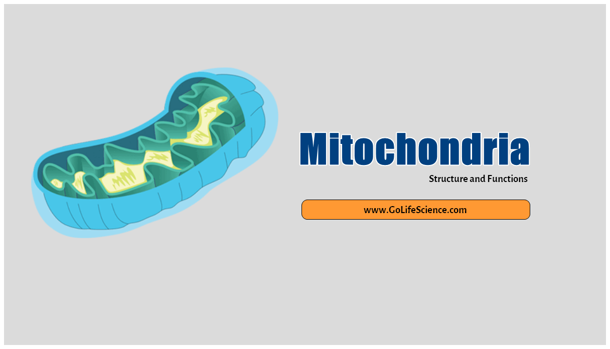
Mitochondrial are present in all eukaryotic cells and are the major sites of aerobic respiration within cells. they were first seen as granules in muscle cells in 1850. The number of mitochondrial per cell varies considerably and depends on the type of organism and the nature of the cell. This article gives the information on Structure and function of mitochondrial.
Historical background:
- 1850 – Kolliker – observed in muscle cells of insects
- 1882 – Flemming– gave the name as “filia”
- 1892 – Altmann– gave systematic name observation name as “Bioblast”
- 1897-98 – Benda– gave a name as “Mitochondrial”. He stained the mitochondrial with “alizarin” and “crystal violet”.
- 1900 – Michaelis– stained mitochondrial with “Janus green”
- 1934 – Bensley & Hopper – said mitochondrial is the site for the cellular respiration.
- 1940- Palade and Sjostrand – They worked out the fine structure of mitochondrial under the electron microscope.
Other name:
- Fuchsinophilic granules,
- Parabasal bodies,
- Plasmosomes,
- Fila,
- Chondriosomes,
- Vernicules
- Bioblasts.
Structure and function of mitochondria

Biochemistry and Anatomy of Mitochondria
Mitochondrial (Mc) was first observed by “Altmann” in 1894 who described as “bioblasts”. Benda (1897) called them “mitochondrial”. (mitoG =thread; chondrionG=granule). Here are the Structure and function of mitochondrial.
- The number of Mc varies with the cell type and functional stages. In eukaryotes, approximately 2000 Mc copies one-fifth of its total cell volume.
- The Mitochondrial’s chemical composition is concerned, It consists of 65-70% proteins, 25-30% lipids, 5-7% mitochondrial DNA and 0.5%RNA. The outer membrane of the mitochondrial has “porins”, which permits molecules up to 10kd.
- Mitochondrial Matrix is gel like a solution, containing the high concentration of soluble enzymes, substrate, nucleotide cofactors, ions.
- The Mc is a subcellular organelle having the outer and inner membranes enclosing the matrix.
- The inner membrane is highly selective in its permeable characteristics.
- The inner membrane contains the respiratory chain and translocation systems.
- The knobs like protrusions represent the ATP synthase system.
- The inner membrane is folded into a series of internal ridges called “Cristae”, which may be longitudinally or transversely oriented, branched or tabular.
Hence, there are two compartments in Mitochondrial: the intermembrane space between the outer and inner membranes and the matrix, which is bounded by the inner membrane. Most of the reactions of the TCA cycle (citric acid cycle ) and fatty acid oxidation occur in the matrix.
Size: The average length of Mc is 3-4 microns and the average diameter 0.5 to 1.0 micron.
Shape: Mitochondrial vary in shape but are generally filamentous or granular. If a filamentous mitochondrion swells at one end, it gives a club shape club-shaped. If the swollen and hollows out then it becomes tennis racket shaped. If there is a central clear zone, the mitochondrion becomes vesicular. According to Green, they are 1500A
Number: The number of mitochondrial varies in different cell types. the number of mitochondrial depends upon the metabolic activity of the cell. in a normal liver cell, there are 1000 to 1600 mitochondrial while in plant cells their number is very small.
Distribution: Ordinarily, The Mc is evenly distributed in the Cytoplasm. In columnar or prismatic cells, Mc is oriented parallel to the long axis of the cell.
Enzymes localization in Mitochondrial
LOCALISATION OF SOME ENZYMES IN RAT-LIVER MITOCHONDRIAL
- Monoamine oxidase
- Kynurenine-3-monoxygenase
- NADH dehydrogenase
- Acyl. CoA Synthetase
- Phospholipase-A2
- Nucleoside diphosphate kinase
- NADPH dehydrogenase
- Iron-Sulfur Proteins
- Cyt.b,c,c1and aa3
- F1 ATPase
- Succinate dehydrogenase
- Carnitine acyltransferase
- TCA Cycle enzymes
- Fatty acyl-CoA oxidation enzyme
- Adenylate kinase
- Creatine Kinase
Chemical Composition of Mitochondrial
- The major constituent of mitochondrial is proteins (4/5) and liquids (1/5).
- The lipid content is composed of 90 percent phospholipids (Lecithin and Cepahlin), about 5 percent free fatty acids, and triacylglycerides.
- Mitochondrial are rich in enzymes related to substrate oxidation, respiration, energy conservation, oxidative phosphorylation, and Kreb’s cycle.
- A heterogeneous group of enzymes related to phospholipid metabolism and fatty acid oxidation is present in the outer compartment and outer membrane.
- Cu++, Mg++, HPO4-, Cl-, RNA, and DNA (without histone, a protein) are also reported to occur.
Functions
What are the functions For Mitochondrial? The Mitochondrion plays an important role in Cellular Respiration.
- The Mitochondrial are organelles that transfer the chemical energy of the metabolites of the cell (through the Krebs cycle and the respiratory chain) into the high-energy phosphate bond of ATP (production of ATP).
- Thus, mitochondrial is the “powerhouse of the cell”, that produces the energy necessary for many vital cellular functions via, motility contraction (muscle contraction), biosynthesis of cell bioluminescence, etc.
