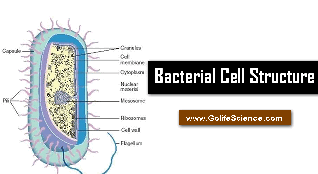
Bacteriology is the study of bacteria. Bacteria are minute, microscopic, simple, unicellular prokaryotic organisms occurring as saprophytes and parasites on a wide range of habitats.

Structurally a bacteria cell consists of three categories of structurally namely,
- Structures of the External side of the cell wall.
- Cell wall
- Structures of the Internal side of the cell wall
External Structures
The bacterial cell Structures at the external side of the cell wall include flagella, fimbriae (pili), and capsule (slime layer).
i) Flagella
Flagella are thin, hair-like appendages that originate from a granular structure, the basal body which is present just beneath the plasma membrane. They are composed of protein, “Flagellin”. The flagellin molecules are forming a single cylindrical filament.

Flagella are present in eubacteria. The bacilli and possess flagella. The cocci do not possess flagella. They are responsible for the motility of bacteria.
On the basis of the arrangement of flagella, four types of arrangements are recognized. They are “Lophotrichous, Amphitrichous, Monotrichous, Peritrichous” (In shortcut for easy recollection- LAAMP).
- Lophotrichous: A tuft of flagella attached to one side of the cell.
- Atrichous : Flagella are totally absent
- Amphitrichous: Two tufts of flagella (or) single flagellum on either end of the cell.
- Monotrichous : A single flagellum is found on one side of the cell.
- Peritrichous : Many flagella found all over the cell surface.
Structurally a flagellum is composed of three parts:
- Basal body
- Hook
- Filament

a) Basal body
It is the most complex part of the flagellum. The basal body is located entirely within the cell envelope and measures about 27nm in length. The basal body consists of a small central rod inserted into a system of rings. The system of rings, however, differs between gram +ve and gram-ve types of Bacteria.
The outer pair called “L” and “P” rings are situated at the level of the outer membrane and serve as bushings for the insertion of the body through the outer membrane.
The inner pair namely “S” and “M” rings are located near the level of the cell membrane.
But in gram +ve Bacteria, the basal body bears only one pair of rings namely “S” and “M” ring. The “L” and “P” rings are absent. The basal body acts as a motor and imparts rotatory motion to the flagellum. The two inner “S” and “M” rings are believed to be responsible for the rotatory motion in gram –ve and gram +ve bacteria.
b) Hook
It is the part that lies between the filament and basal body of the flagellum. The hook is slightly wider than filament and is about 45nm in length. It is made up of protein subunits, which are antigenically different from that of flagellin sub-units.
c) Filament
The outermost structure of the flagellum is the filament. The filament is a helical structure with a 14nm diameter. It is composed of subunits or monomers of a protein known as flagellin.

The molecular weight of the flagellin is about 40,000. The shape of the flagellum is dependent on flagellin.
The flagellum grows at its rather than at the base as the newly synthesized flagellin monomers are added at the distal end of the filament.
Hydrodynamics of flagella:
Bacteria propel themselves by rotating their helical flagella. The principle involved can be illustrated by imaging the penetration of a piece of cork by a corkscrew. In the case of bacteria, the cork is analogous to the viscous medium and the corkscrew to the helical flagellum.
The nature of the rotatory motor that spins each corkscrew-shaped flagellum is still not understood, but the rings found in the basal body are probably involved.
It is known that the flagella derived from the electrical potential and hydrogen ion gradient across the cytoplasmic membrane.
According to recent studies, the concentration of cGMP within the cell governs the direction in which the rotatory occurs.
Swimming motility without flagella
Certain helical bacteria (spirochetes) exhibit swimming motility, particularly in highly viscous media, yet they lack that is flagella.
Inside the bacteria, flagella like structure are present, that is “Axial fibril” (or) “endoflagella” responsible for motility of the spirochetes, the mechanism is not yet clear.
Gliding motility
Some Bacteria (eg: Cytophage species) are motile only when they are in contact with a solid surface. This type of motion comparatively slow (few mm/sec). No special organelles have been observed.
ii) Pilli (Fimbriea)
Many gram negatives Bacteria possess minute, shorter, hair-like hallows appendages called the “Pilli”.
They originate from the cell wall. They are composed of protein, pillin, they are organs of attachment.
The one kind of pili called sex pilus, it serves as the part of the entry of DNA during bacterial conjugation.
Some pili play a major role in human infection by allowing pathogenic bacteria to attach to epithelial cells lining the respiratory, intestinal or genitourinary tracts.
iii) Capsule
A viscous layer called the slime layer (or) capsule surrounds some Bacterial cells. The capsule is secreted by the cytoplasm and contains polysaccharides or peptides.. it protects the bacteria and may serve as a reservoir of stored food. Or waste products. Capsulated bacteria are infectious while non-capsulated Bacteria are noninfectious.
The main functions of the capsule or slime layer are
- It may provide protection against temporary drying by binding water molecules.
- They may block the attachment of bacteriophages.
- They may be antiphagocytic i.e., they inhibit the engulfment of pathogenic bacteria by WBC and thus contribute o invasive or infective ability (virulence).
- They may promote attachment of Bacteria to surfaces.
Most Bacterial capsules are composed of polysaccharides, few capsules are polypeptides.
Capsules composed of a single kind of sugar are termed homopolysaccharides, several kinds of sugars are termed as heteropolysaccharides.
Bacterial Cell Wall
The bacterial cell wall is surrounded by a rigid cell wall.
The cell wall is one of the most important layers of the prokaryotic cell.
In bacteria generally, the layer or layers or layer of cell envelope lying between the inner cytoplasmic membrane and the capsule is called “Cell wall”.
The main functions of the cell wall are:
- It gives shape and rigidity to the cell.
- It provides solid anchoring support to flagella.
- It prevents the lysis or rupture of Bacteria due to osmotic pressure.
- It contributes to the pathogenicity of many pathogens.
- It can protect a cell from toxic substances and is the site of action of several antibiotics.
Under the external structure as capsules sheaths and flagella and external to the cytoplasmic membrane is the “cell wall”, a very rigid structure that gives shape to the cell.
Its main function is to prevent the cell from expanding and eventually bursting because of the uptake of water since most Bacteria live in a hypotonic environment.
The walls of gram -ve species are generally thinner (10 to 15 nm) than those of gram +ve species (20 to 25 nm). The walls of gram –ve archebacteria are also thinner than those of gram +ve archebacteria.
Chemical composition
For eubacteria, the shape- determining part of the cell wall is large “Peptidoglycan” (sometimes called –“MUREIN”), an insoluble porous, cross-linked polymer of enormous strength and rigidity.
The peptidoglycan is found only in prokaryotes, it occurs in the form of a “Bag shaped macromolecule” surrounding the cytoplasmic membrane.
The peptidoglycan basically a polymer of:
- N- Acetylglucosamine,
- N- Acetylmuramic acid,
- L-Alanine,
- D- Alanine,
- D-Glumatic acid and
- Diamino acid
(LL – (or) meso = diamino pimelic acid, L-Lysin, L-Ornithine, (or) L-diamino butyric acid
In archaebacteria possess cell walls, these do not contain peptidoglycan, and their cell wall fine structure and chemical composition are very different from that of eubacteria.
Their cell walls are usually composed of proteins, glycoproteins (or) Polysaccharides.
- Methanobacterium have cell walls –Pseudomurein”
Bacteria are divided into 2 types, namely gram +ve and gram –ve basing o the differences in the reaction to gram’s staining.
Walls of gram – Positive eubacteria
Gram +ve bacteria usually have a much great amount of peptidoglycan in their cell walls than do gram –ve bacteria (~50% (or) more of the dry weight), but only in about 10% of the wall of gram-negative bacteria.
Other substances may occur in addition to Peptidoglycan.
- Streptococcus pyrogens contain “Polysaccharides”, which can be extracted with hot-dilute HCl.
- “Streptococcus aureus” and “Streptococcus faecalies” contain ‘Teichoic acid”– acidic polymers of ribitol phosphate (or) glycerol phosphate, which can be extracted with cold dilute acid.
- Teichoic acid binds Mg+2 ions and there is some evidence that they help to protect Bacteria from thermal injury by providing an accessible pool of these cations for stabilization of the cytoplasmic membrane.
- The walls of the Bacteria can be easily destroyed by treatment with an enzyme called “Lysozyme”, which selectively dissolves Peptidoglycan.
Walls of gram – Negative eubacteria
The walls of gram-negative bacteria are more complexes than those of gram-positive bacteria.
The most interesting difference in the presence of an ‘outer membrane’ that surrounds a thin underlying layer of Peptidoglycan.
The walls of gram-negative Bacteria are rich in lipids (11 to 22% dry weight of the wall).
- This outer membrane serves as an impermeable barrier to prevent the escape of important enzymes, such as those involved in cell wall growth from the space between the cytoplasmic
- Gram –ve Bacteria are refractory to the enzyme “Lysozyme” (or) resistant to the lysosomal treatment.
- Only if the outer membrane is first damaged, as by removal of stabilizing “Magnesium ions” by a chelating agent, can the enzyme penetrate and attack the underlying Peptidoglycan layer?
- The outer membrane of the gram –ve cell wall is anchored to the underlying Peptidoglycan by means of “Braun’s lipopolysaccharides”.
- The membrane is a bi-layered structure consisting mainly of “Phospholipids, proteins, and lipopolysaccharides (LPS)”.
- The LPS has toxic properties and is also known as “Endotoxin”. It composed of three covalently linked pats.
- a) Lipid.A: Firmly embedded in the membrane.
- b) Core polysaccharide: Located at the membrane surface.
- c) O-linked polysaccharide: which extend like whiskers from the membrane surface into the surrounding medium. Many of the serological properties of gram-negative Bacteria are attributable to O-antigens; they can also serve as receptors for Bacteriophage attachment.
Internal Structures
The structures that present interior to the cell wall include cell membrane, mesosomes, cytoplasm, nuclear material, cytoplasmic inclusions, and vacuoles.
i) The cell membrane (or) Cytoplasmic membrane
Immediately beneath the cell wall, there is a thin membrane around the cytoplasm.
This membrane is called the cytoplasmic membrane or plasma membrane. It contains phospholipids, proteins, and polysaccharides.
The cell membrane is a vital structure and critical barrier that separates the inside of the cell from the outer environment.
The most widely accepted current model for cell membrane structure is the fluid mosaic proposed by Singer and Nicholson.
The thickness of the cell membrane is 7.5nm. the plasma membrane is semi-permeable and regulates the transport of nutrients and waste products into and out of the cell.
The cell membrane is composed of 20 – 80% of phospholipids and 60 to 70% of proteins.
The phospholipids are amphipathic and from a lipid bilayer with a hydrophobic group (fatty acid) towards inside and hydrophilic groups towards outside.
Proteins are embedded in the lipid matrix and are called Integral or intrinsic proteins (70 to 80%). 20 to 30% of the protein are loosely connected to the membrane are called peripheral or extrinsic proteins.
Functions:
- selective permeability and transport of solutes.
- Electron transport and oxidative phosphorylation in aerobic species.
- Excretion of hydrolytic exoenzymes.
- Bearing the enzymes and carrier molecules that function in the biosynthesis of DNA, cell wall polymer and membrane lipids.
- Bearing the receptors and other proteins of the chemotactic and other sensory transduction systems.
ii) Mesosomes
In gram-positive bacteria, the plasma membrane forms in folding.
The infolding can give rise to mesosomes within the cytoplasm.
The mesosome is associated with bacterial nuclear material and its replication. Respiratory enzymes are also associated with thin mesosomes.
Functions:
- They involve in cell wall formation during cell division.
- They play a role in the replication of chromosome and distribution to daughter cells.
- They also involve in secretory processes.
iii) Cytoplasm
The cytoplasm is an aqueous solution of soluble proteins and metabolites.
It contains RNA, ribosomes, and reserve food materials. Is also contains a nuclear body or nucleoid.
iv) Nuclear material
The prokaryotic cell is strikingly different from the eukaryotic cell in the lack of a well-defined nucleus.
The nuclear body or nucleoid lacks a nuclear envelope. It consists of a single molecule of DNA which is circular.
It embedded directly in the cytoplasm. It consists of a nuclear zone at the center of the cell.
This nuclear zone is also called as a nuclear body or chromatin body or nuclear region or nucleoplasm or nucleoid.
In addition to this genetic material (DNA), many bacteria possess other genetic material called Plasmids.
The plasmids are non-chromosomal genetic material. Plasmids are circular, double-stranded DNA molecules with independent replicating capacity, they are smaller in size.
- What are DNA Polymerase and its function in DNA Replication
- What are the Origin of replication in Prokaryotes and Eukaryotes
v) Ribosomes
In the bacterial cells, ribosomes occur freely in the cytoplasm and in clusters of 4 to 6 ribosomes. The clusters are called the polysomes.
The ribosome is of the 70S consisting of 50S and 80S particles. The cytoplasmic area, granular in appearance and rich in the macromolecular RNA protein bodies known as Ribosomes, on which protein is synthesized.
vi) Cytoplasmic inclusions and Vacuoles
Concentrated deposits of certain substances are detectable in the cytoplasm of some bacteria. The reserved food materials are fats, polysaccharides, volutin, etc.
Volutin granules are also known as metachromatic granules are composed of polyphosphate. Volutin serves as a reserve source of phosphate.
Fats are stored in highly retractile globules in the form of polymerized β-hydroxybutyric acid, which can serve as a reserve carbon and energy source polysaccharides (glycogen) are stored in granules.
- The Cell – Structure, and Functions (Synopsis Points)
- Branches of Life Sciences – Brush Back Your Basics
Another type of inclusions is represented by the intracellular globules of elemental sulfur that may accumulate in certain bacteria growing in environments rich in hydrogen sulfite.
Some bacteria that live in aquatic habitats form gas vacuoles that provide buoyancy, these are bright retractile bodies and have a regular shape, hallow, rigid, cylinders with more or less conical ends and having a striated protein boundary. This boundary is impermeable to water.
